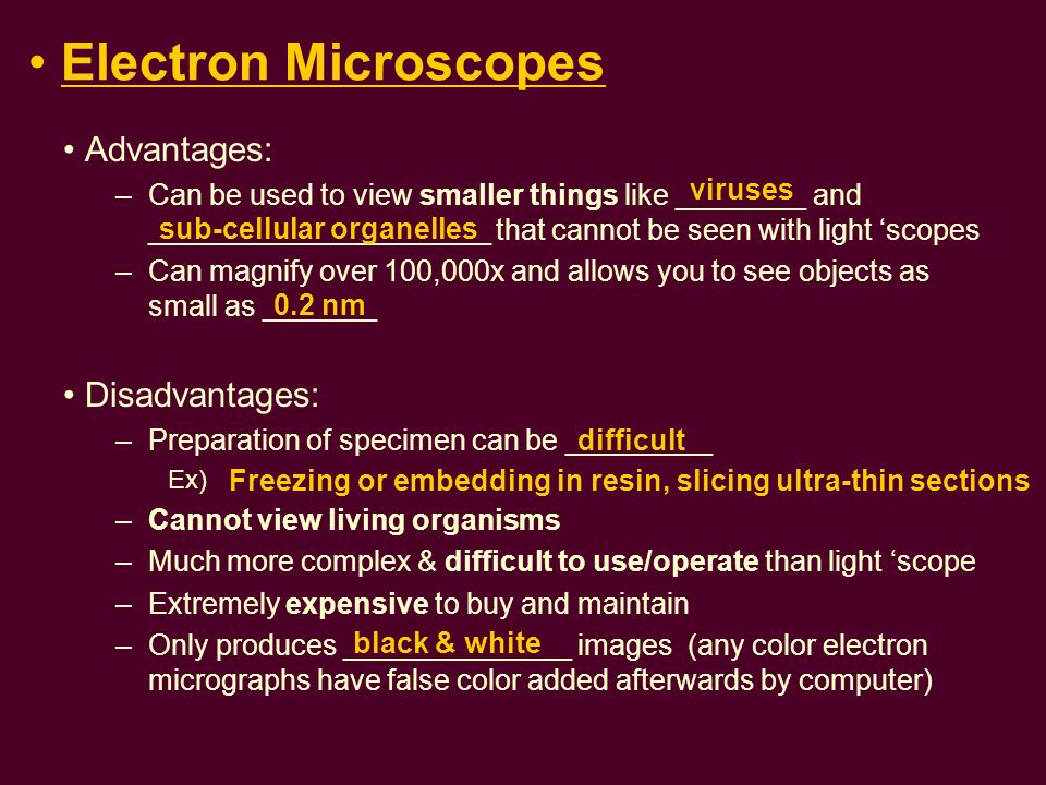
 alt='disadvantages of electron microscope vs light microscope' title='disadvantages of electron microscope vs light microscope' />.Others include back-scattered or reflected electrons and cathodoluminescence.The most commonly used method in biological applications is secondary electron detection from excited atoms within the beam.The beam position is closely linked with the emission signal to construct a resulting image in fine detail - at a resolution in the low nm range.These electrons interact with electrons in the sample and produce signals representative of the sample topography and composition.SEM involves the production of an image by scanning the specimen with a focused beam of electrons. The electron microscope concept overcomes the diffraction limit and permits imaging of much smaller and more complex substances and biological specimens. The lowest diffraction achievable with conventional lenses is 200 nm, and efforts to surpass this point have been limited even with modern techniques, although there are notable exceptions. Consequently, there is a limit (the Diffraction Limit) beyond which the ability to resolve two images becomes impossible.Diffraction (or the mathematically defined Diffraction Limit) is used as the measure of resolution and is impacted by factors including: the wavelength of incident light, the refractive index of materials through which the light passes, and the numerical aperture of the lens used to magnify the image.In other words, the ability to reveal structural details as distinct and separate.

Resolution is perhaps the central tenet of microscopy in that it defines the ability of any device to distinguish between two closely spaced objects.Three main factors underlie the scientific principle of microscopy: magnification, illumination, and resolution.
